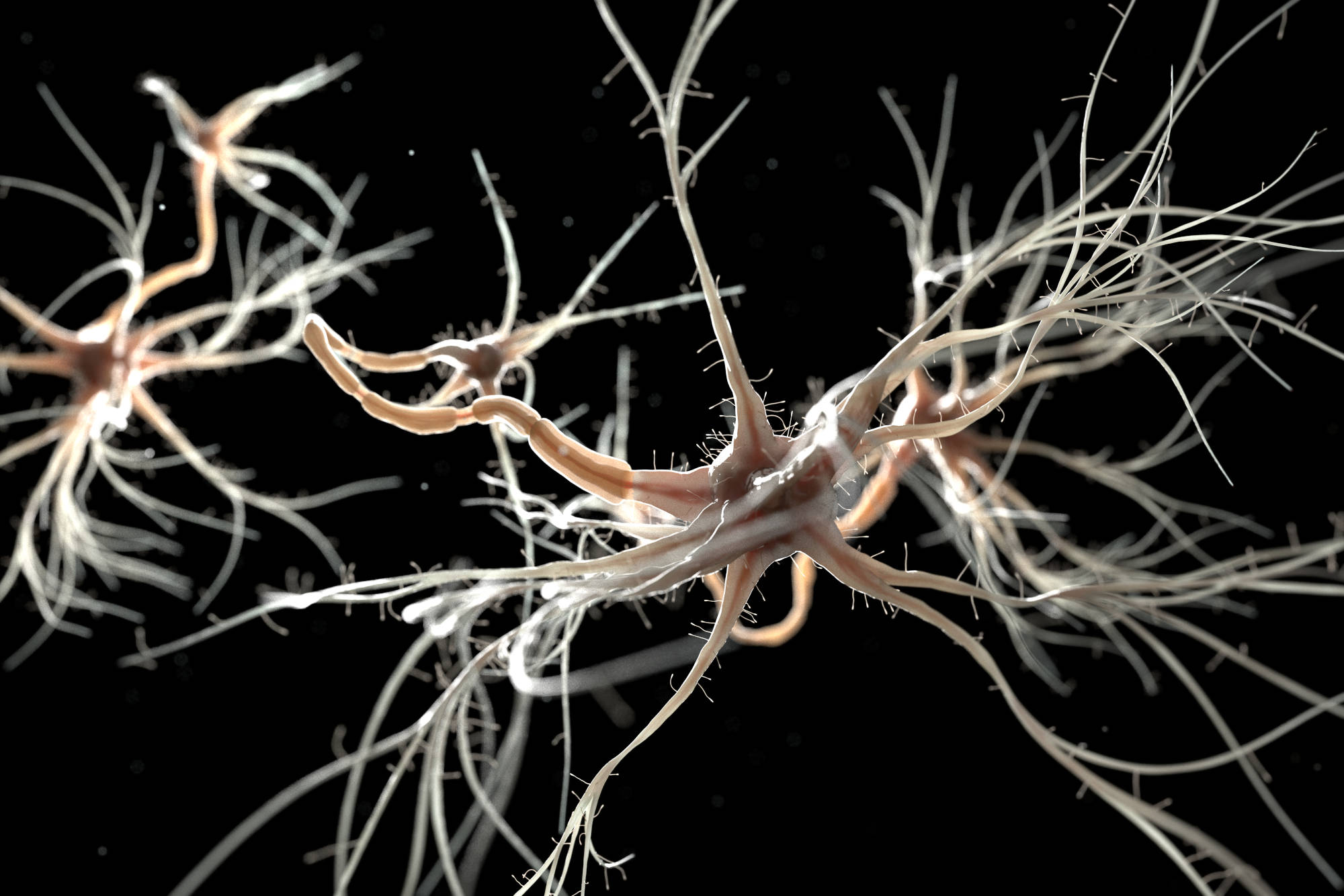
After a peripheral nerve is damaged, the nerve stump tries regenerating in the direction of its distal target [1]. When the nerve regenerates in a disorganized fashion or cannot complete the process at all, a neuroma forms [1]. Neuromas are growths of nerve tissue that can result in psychologically and physically debilitating centrally-located pain [1, 2]. They can be especially painful if the neuroma is shallow or situated near the site of tendon excursion or a joint because, then, the nerve’s axons will be susceptible to frequent stimulation [2]. Accordingly, the symptoms associated with neuromas are sharp pain, numbness, electrical sensitivity, paresthesia, and cold intolerance [1]. Unfortunately, treatment for a neuroma is difficult [2]. An array of treatment options exist, such as neuromodulation, radiofrequency, ablation, desensitization, and pain medications, but none of them are definitively effective [1]. Pharmacotherapy is often ineffective, as are other symptomatic treatments [1]. Neuroma treatment is complicated by the short time during which it must occur to be successful. In 2018, Oliveira et al. studied the development of neuromas over time by assessing the nerve morphology of 30 amputated Sprague Dawley rats [3]. They found that neuroma prevention and treatment options are likely to be much more fruitful if they occur within 28 days of amputation [3]. Having to operate within this compressed timeline may complicate treatment options further [3].
Surgery is a common treatment option. A retrospective study conducted by Stovkis et al. reported that 56% of patients are satisfied after surgery [4]. Not only can mean visual analog scale scores decrease significantly, but upper extremity function also tends to improve greatly [4]. One of the most frequently performed operations is a translocation of the nerve stump into a piece of their muscle tissue [2]. Following this procedure, pain relief occurs at rates ranging from 35% to 100%, suggesting that it can be highly successful in some cases but is not adequately effective enough to become a blanket treatment for all patients [2].
Transposition of the nerve stump into a nearby vein, rather than muscle tissue, has emerged as an alternate procedure for neuromas [2]. The results of this procedure are promising: one study reported that recipients of this operation experienced less intense pain and improved evaluative, affective, and sensory dimensions of pain in comparison to patients who underwent nerve stump translocation [2]. Furthermore, transposition patients reported improved upper extremity function and the ability to partake in more physical activity [2]. Another study, conducted by Koch et al., had similarly promising results. Neuroma patients who received transpositions experienced significant symptom relief, with 12 of the 23 patients cured of their neuroma-induced pain [5]. While neither experiment indicates that transposition is successful in all cases, it seems preferable to translocation in these instances.
Another form of neuroma treatment is the implantation of regenerative peripheral nerve interfaces [6]. RPNIs are muscle grafts that prevent neuroma formation by serving as physiological targets for incoming peripheral nerve growth [6]. Amputees who suffer from symptomatic neuromas can receive RPNIs to relieve their pain [6]. In 2016, Woo et al. conducted a retrospective analysis of 16 RPNI recipients [6]. They found that 71% of subjects reported reduced neuroma pain, and 53% experienced diminished phantom pain [6]. As a result of their RPNIs, the patients also used either less or more stable amounts of analgesics [6]. While complications of the procedure included pain at a different bodily region or slowed wound healing, this study presents a promising additional option for physicians treating neuroma patients [6].
Despite the difficulties of neuroma treatment, these techniques suggest that there are many ways to alleviate patients of the chronic pain associated with neuroma so that they can lead unhindered lives.
References
[1] K. R. Eberlin and I. Ducic, “Surgical Algorithm for Neuroma Management: A Changing Treatment Paradigm,” Plastic Reconstructive Surgery Global Open, vol. 6, no. 10, p. 1-8, October 2018. [Online]. Available: https://doi.org/10.1097/GOX.0000000000001952.
[2] H. Balcin et al., “A comparative study of two method of surgical treatment for painful neuroma,” The Journal of Bone and Joint Surgery, vol. 91-B, no. 6, p. 803-808, June 2009. [Online]. Available: https://doi.org/10.1302/0301-620X.91B6.22145.
[3] K. M. C. Oliveira et al., “Time course of traumatic neuroma development,” PLOS ONE, vol. 13, no. 7, p. 1-15, July 2018. [Online]. Available: https://doi.org/10.1371/journal.pone.0200548.
[4] A. Stokvis et al., “Surgical management of neuroma pain: A prospective follow-up study,” PAIN, vol. 151, no. 3, p. 862-869, December 2010. [Online]. Available: https://doi.org/10.1016/j.pain.2010.09.032.
[5] H. Koch et al., “Treatment of Painful Neuroma by Resection and Nerve Stump Transplantation Into a Vein,” Annals of Plastic Surgery, vol. 51, no. 1, p. 45-50, July 2003. [Online]. Available: https://doi.org/10.1097/01.SAP.0000054187.72439.57.
[6] S. L. Woo et al., “Regenerative Peripheral Nerve Interfaces for the Treatment of Postamputation Neuroma Pain: A Pilot Study,” Plastic Reconstructive Surgery Global Open, vol. 4, no. 12, p. 1-8, December 2017. [Online]. Available: https://doi.org/10.1097/GOX.0000000000001038.
