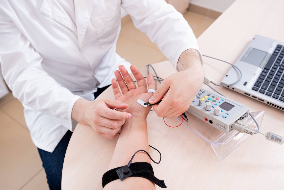
A relatively new diagnostic tool, electrodiagnostic testing is used to investigate the electrical activity of nerves and muscles, especially when a patient seeks treatment for pain with an unclear source [1]. While the most common electrodiagnostic tests — electroencephalograms (EEG) and electrocardiograms (ECG or EKG) — are utilized in the contexts of specific conditions and routine care, other forms of electrodiagnostics can often help determine the cause and extent of a muscle or nerve pain, weakness, or numbness, and can also be used to evaluate the response of a damaged nerve or muscle to pain management treatment [1]. Based on the results of electrodiagnostics, conditions causing pain, such as carpal tunnel syndrome, herniated discs, and amyotrophic lateral sclerosis (ALS), can be diagnosed [2]. When prescribed to evaluate the cause and extent of pain, electrodiagnostics typically includes two separate tests: a nerve conduction study (NCS) and electromyography (EMG) [1, 2]. Although these tests are usually not the sole diagnostic methods utilized to establish a diagnosis for pain and are often referred to as an extension of the physical examination, they reveal essential information about the cause of a suspected myopathy that cannot be determined otherwise [3, 4, 5].
While the typical electrodiagnostic workup includes two similar tests, each of them analyzes a separate component of the body in order to determine whether the symptoms have arisen from nerve damage or a muscle injury. Both tests involve electric currents administered to parts of the body, but potential complications are minimal [6]. The first element of the workup, a nerve conduction study (NCS), administers small electrical currents through electrodes placed on the surface of the skin to measure the speed and amplitudes of the electrical activity of both sensory and motor nerve fibers [1]. Abnormalities recorded by an NCS, such as a low velocity of electrical conduction in a nerve, indicate that nerve damage is responsible for or at least involved in the patient’s symptoms [2]. The second element of the workup, electromyography (EMG), is commonly known as “needle EMG” because it uses needle electrodes inserted into the muscle to record the electrical activity of muscle fibers during certain movements [2]. Abnormal EMG results indicate muscle dysfunction, which can be caused by muscle damage or an underlying nerve problem [3]. Although both NCS and EMG provide excellent information about the suspected condition, experts agree that these tests are not conclusive enough to be used in the absence of other testing, a patient history, and imaging [3, 4, 5].
While informative, electrodiagnostics only compose one part of the diagnostic process. Other components, such as magnetic resonance imaging (MRI), x-ray, and patient history, are essential to create a complete understanding of the patient’s condition. However, studies have shown that electrodiagnostic testing is able to identify and specify the cause and extent of certain myopathies in MRI-negative patients [3, 4]. Moreover, in some cases, results of electrodiagnostics are less likely to be falsely positive compared to MRI results [5]. Thus, electrodiagnostics constitute an important element of the diagnostic process, especially when routine diagnostic testing results are inconclusive, but patient history, MRI, x-ray, and other diagnostic tests cannot be overlooked.
References
1: Bowen, J. (2009). “Chapter 47 – Electrodiagnostic testing” in The Sports Medicine Resource Manual, pp. 568-573. DOI: 10.1016/B978-141603197-0.10046-1.
2: Feinberg, J. (2009). EMG testing: a patient’s guide. Harvard Hospital for Special Surgery. Online article. URL: https://www.hss.edu/conditions_emg-testing-a-patient-guide.asp.
3: Hasankhani, E. and Omidi-Kashani, F. (2013). Magnetic resonance imaging versus electrophysiologic tests in clinical diagnosis of lower extremity radicular pain. ISRN Neuroscience, vol. 2013. DOI: 10.1155/2013/952570.
4: Haig, A., Tong, H., Yamakawa, K., Quint, D., Hoff, J., Chiodo, A., Miner, J., Choksi, V., Geisser, M., and Parres, C. (2006). Spinal stenosis, back pain, or no symptoms at all? A masked study comparing radiologic and electrodiagnostic diagnoses to the clinical impression. Archives of Physical Medicine and Rehabilitation, vol. 87. DOI: 10.1016/j.apmr.2006.03.016.
5: Chiodo, A., Haig, A., Yamakawa, K., Quint, D., Tong, H., and Choksi, V. (2007). Needle EMG has a lower false positive rate than MRI in asymptomatic older adults being evaluated for lumbar spinal stenosis. Clinical Neurophysiology, vol. 118. DOI: 10.1016/j.clinph.2006.12.004.
6: Ellison, D., Williams, M., Moody, G., and Farrar, F. (2010). Electrodiagnostic studies. Critical Care Nursing Clinics of North America, vol. 22. DOI: 10.1016/j.ccell.2009.10.011.
